




Stereotactic Ablative Radiation Therapy (SABR)
SABR aims to specifically target tumours in the body with high doses of radiation therapy, while limiting the dose to surrounding healthy organs.1,2
Additional information about SABR as a treatment option includes:
- In some instances and dependent upon tumour type and size, SABR may be considered for treatment rather than surgery1,3
- SABR is used to treat a range of cancer types. The treating doctor will be able to provide further details about which tumour type, and how these are treated using SABR1
- Your treating doctor and care team will discuss this with you
- The number of treatment sessions you require will depend on your individual circumstances1
Image guidance for SABR
The aim of SABR is to minimise the radiation dose to healthy surrounding tissue, which helps to reduce the risks of potential side effects.2 SABR uses a higher dose of radiation compared with standard external beam radiation therapy, delivered directly to the target area, leaving smaller margins (area surrounding the target that may also be affected). It requires millimetre accuracy so relies on imaging to make sure the target is hit directly every time.4
Technologies for image guidance in SABR include:4
Stereoscopic X-ray imaging (the technology used for CyberKnife)27
You can only see high-density objects like bones and any fiducial markers inserted, such as the gold seed fiducial markers used for some prostate cancers. In this image you can see the gold seed fiducial markers in white.
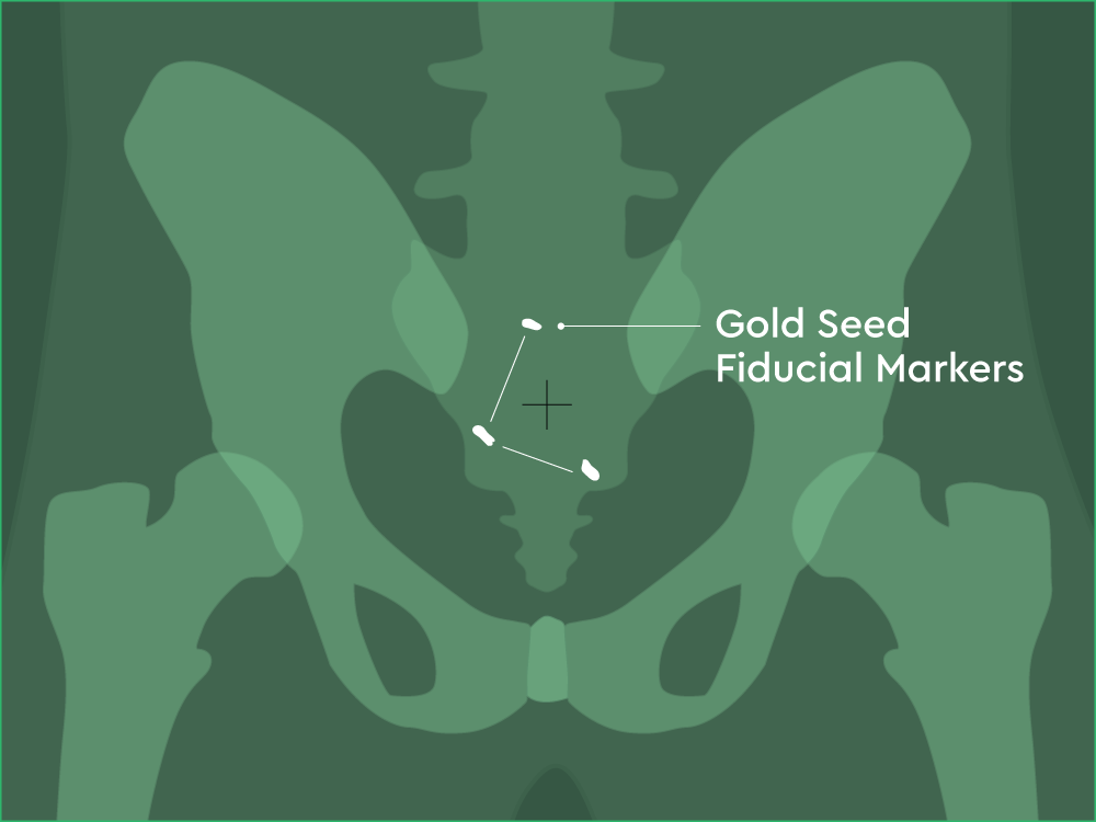
Image A: Front view

Image B: Clinical imaging scan
With this technology you can see more than with stereoscopic X-ray imaging. You can see the same high-density objects as above (bones and fiducial markers), but you can also see more details, such as soft tissues, in three dimensions.
In images A, B and C, you can see the:
- Prostate in red
- Bladder in orange
- Planned target volume outlined in yellow (where the radiation will hit)
In image D, you can see the:
- XXX outlined in blue
- YYY outlined in orange
This level of imaging is standard at GenesisCare. Please note that some SABR treatments cannot be perfomed with CBCT.
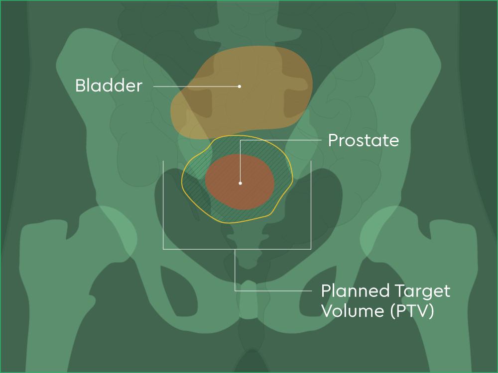
Image A: Front view
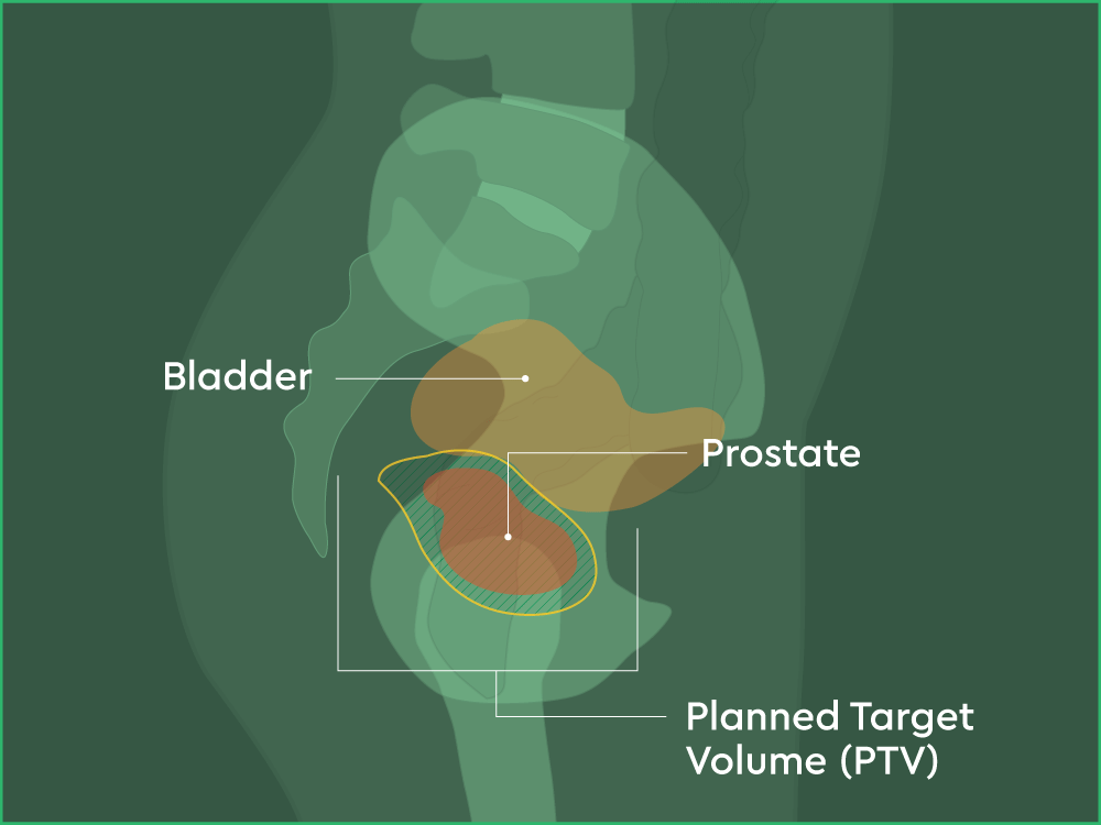
Image B: Side View
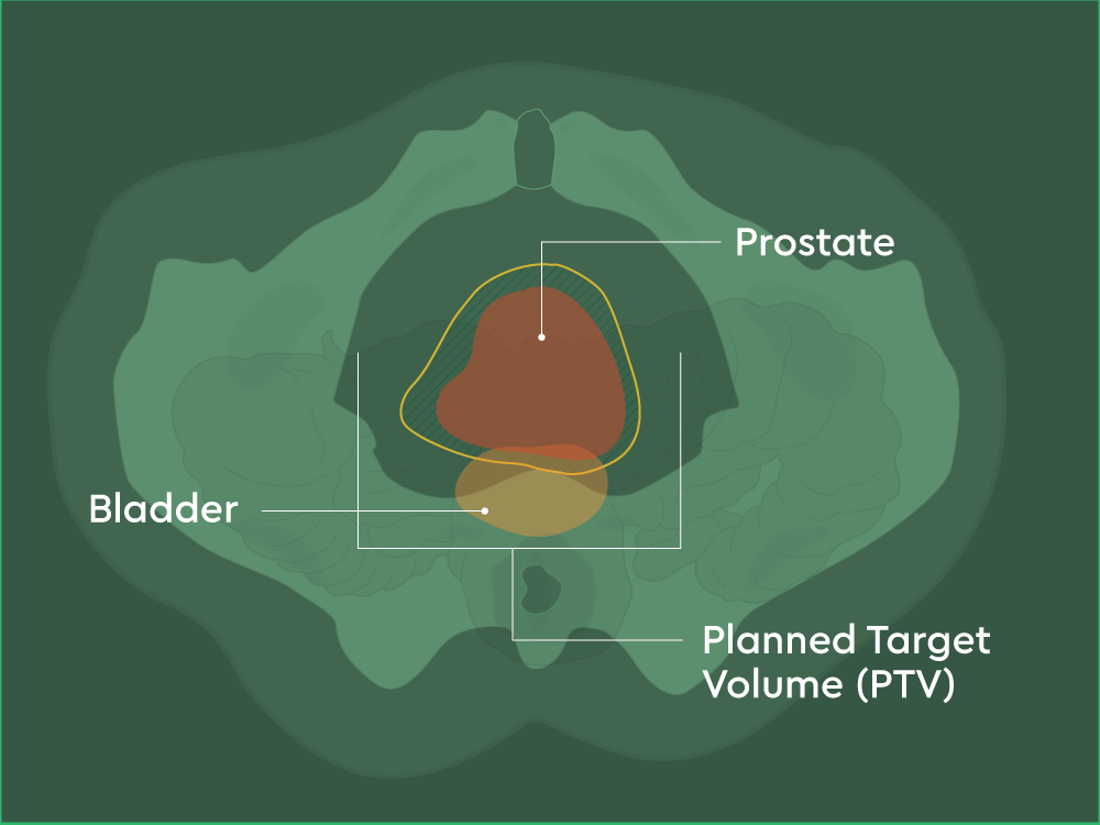
Image C: Bottom View
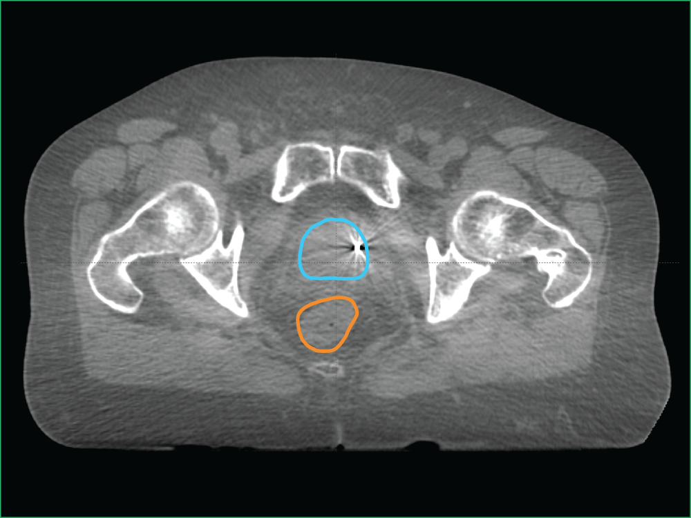
Image D: Clinical imaging scan
Here you can see the high-density objects but also the soft tissue in greater detail in three dimensions, thanks to the MRI.
In image A, you can see the:
- Prostate in red
- Bladder in orange
- Planned target volume outlined in yellow (where the radiation will hit)
In image B, you can see the:
- XXX outlined in blue
- YYY outlined in orange
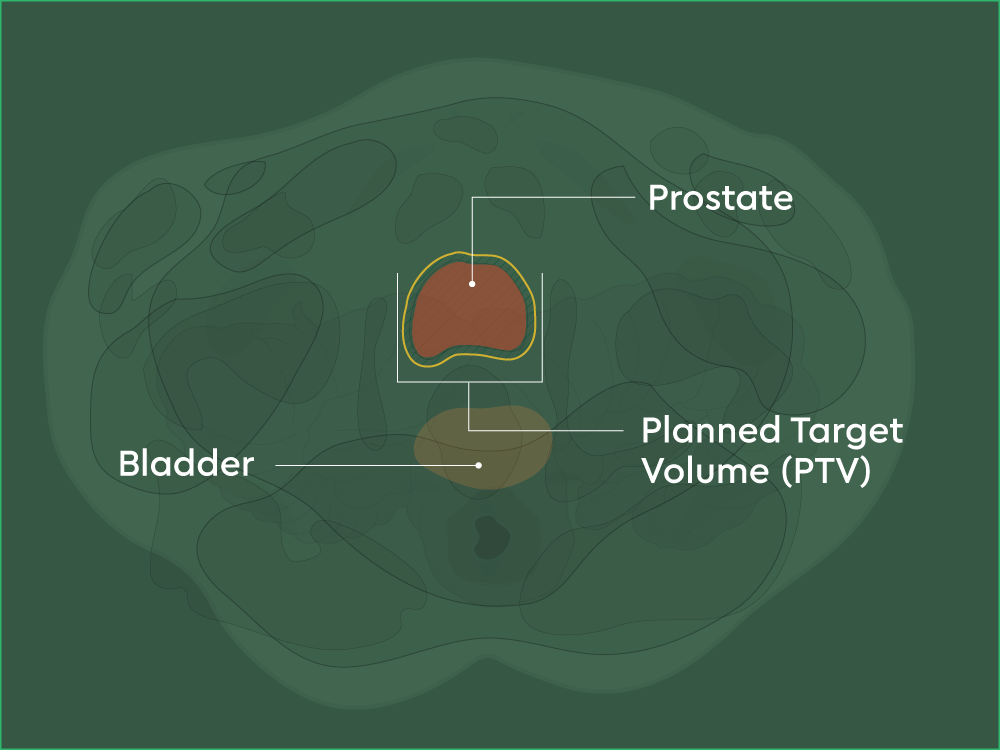
Image A: Bottom view
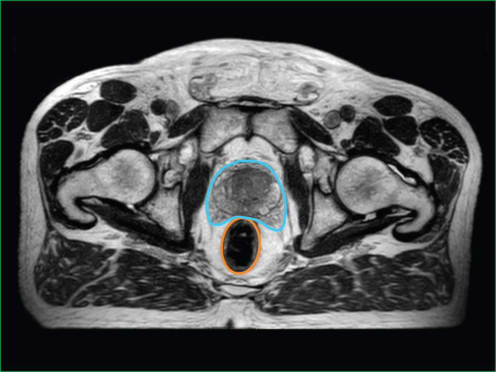
Image B: Clinical imaging scan
Find a centre near you
Read next

Our centres
In Australia, we have more than 40 oncology centres in metro and regional Queensland, New South Wales, Victoria, South Australia, and Western Australia.

Our doctors
Our experienced, specialised doctors offer bespoke, dedicated care aiming to provide the best possible clinical outcomes.

Radiation therapy for cancer
Radiation therapy uses high energy X-rays or other particles to treat cancer and can be used at all stages.
- Cancer Council. Understanding Radiation Therapy. Available at: https://www.cancer.org.au/assets/pdf/understanding-radiation-therapy-booklet (accessed June 2025).
- Tsang Maverick WK. Journal of Thoracic Disease 2016;8:S517–S527.
- Andruska N et al. Journal of Thoracic Oncology 2021;16:1075–85.
- Radiation Oncology Targeting Cancer. Stereotactic Ablative Radiotherapy in AU and NZ. Available from: https://www.targetingcancer.com.au/radiation-therapy/stereotactic-ablative-radiotherapy-sabr/#:~:text=SABR%20uses%20Xray%20beams%20directed,%2C%20Cyberknife%2C%20Tomotherapy%20and%20Gammaknife. (accessed June 2025).
- Kurup G. J Med Phys 2010;35(2):62–64.
- Hall WA, et al. CA Cancer J Clin 2022;72:34–56.
- RadiologyInfo.org. Image-guided Radiation Therapy (IGRT) Available from: https://www.radiologyinfo.org/en/info/igrt (accessed June 2025).